Meet the Scutoid! It's Part of Your Body
In 2018, scientists discovered a new 3D geometric shape called the scutoid — a strange hybrid of a prism and a pyramid that epithelial cells use to efficiently pack curved tissue surfaces in living organisms.
Hi, my name is Diana, I'm a mathematician and I write for the MTS Habr blog. My previous post was about the Borsuk-Ulam Theorem, and today I want to tell you about a discovery from 2018 that sits at the intersection of mathematics and biology.
This article is about a three-dimensional shape — the scutoid — that allows cells to compactly fill curved space.
Where Biologists Found the Scutoid and Why It's Called That
In 2018, a Spanish-American research team studied epithelial cells using confocal microscopy and 3D reconstruction. They examined tissues including Drosophila salivary glands, embryos, and egg chambers.
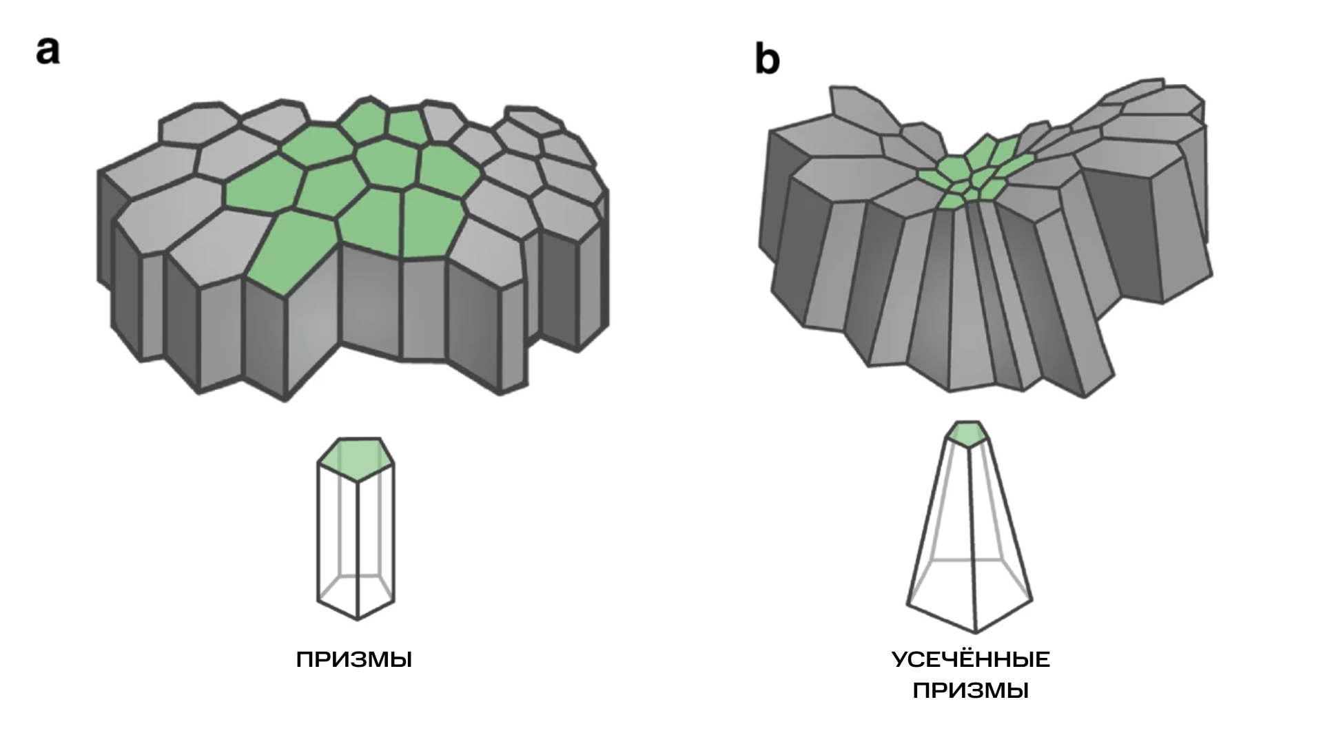
The key discovery: Scientists had previously assumed that cells took the form of prisms or truncated pyramids. However, when tissue curves, cells assume a new shape — especially when the top and bottom layers undergo different degrees of stretching.
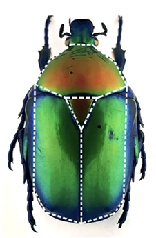
Origin of the name:
- The shape resembles the dorsal shield (scutellum) of beetles (Latin scutum = "shield")
- The lead biologist was Luis M. Escudero — the shape was jokingly called "escu-toid," and the name stuck
What Is a Scutoid from a Mathematical Perspective
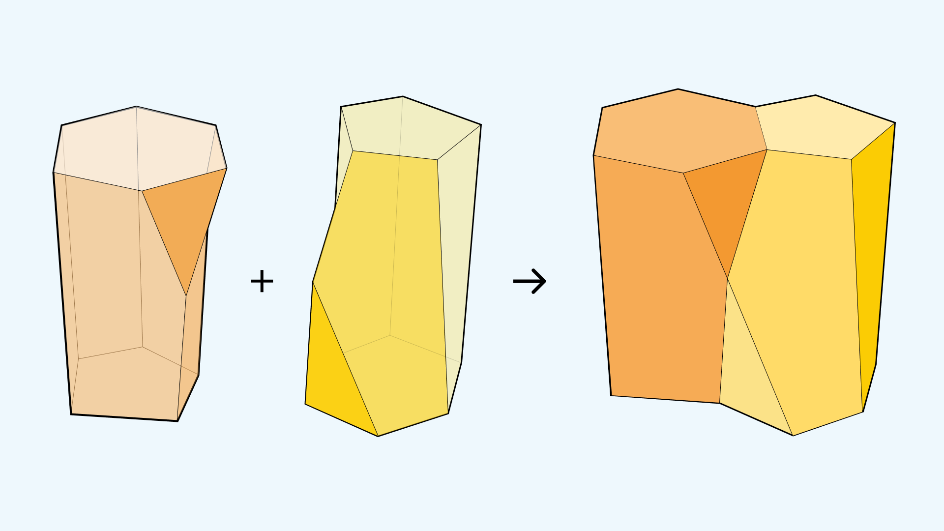
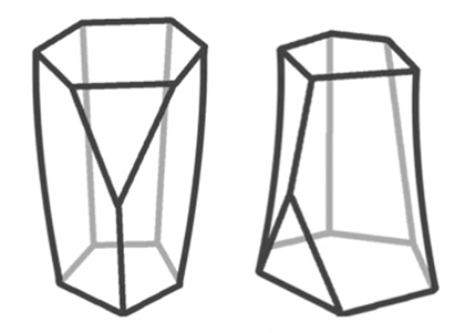
Definition: A scutoid is a strange offspring of a prism and a pyramid.
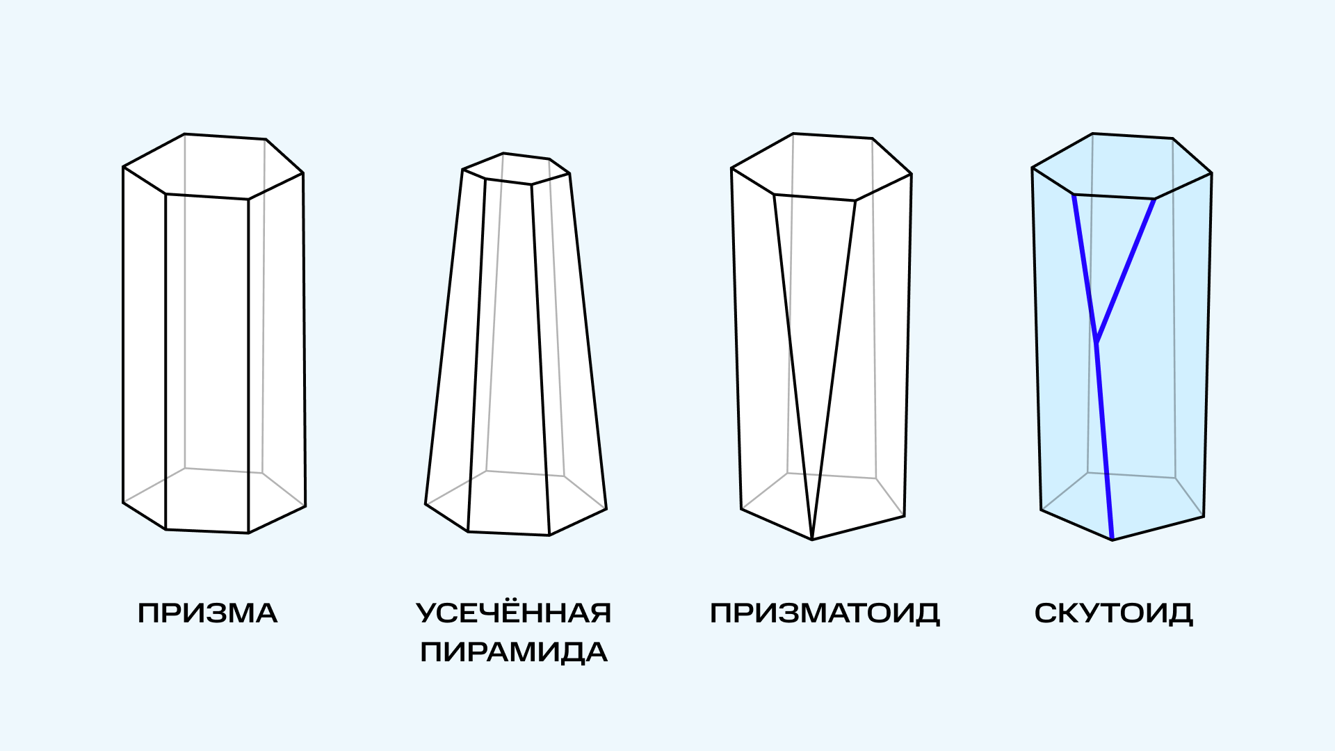
Construction:
- Parallel bases (for example, a pentagon and a hexagon)
- Since the top has more vertices, two upper vertices connect to one lower vertex in a Y-shaped manner
- An additional vertex on the side is required (a mandatory condition)
Key feature: A scutoid is not a polyhedron — its lateral faces are curved rather than flat.
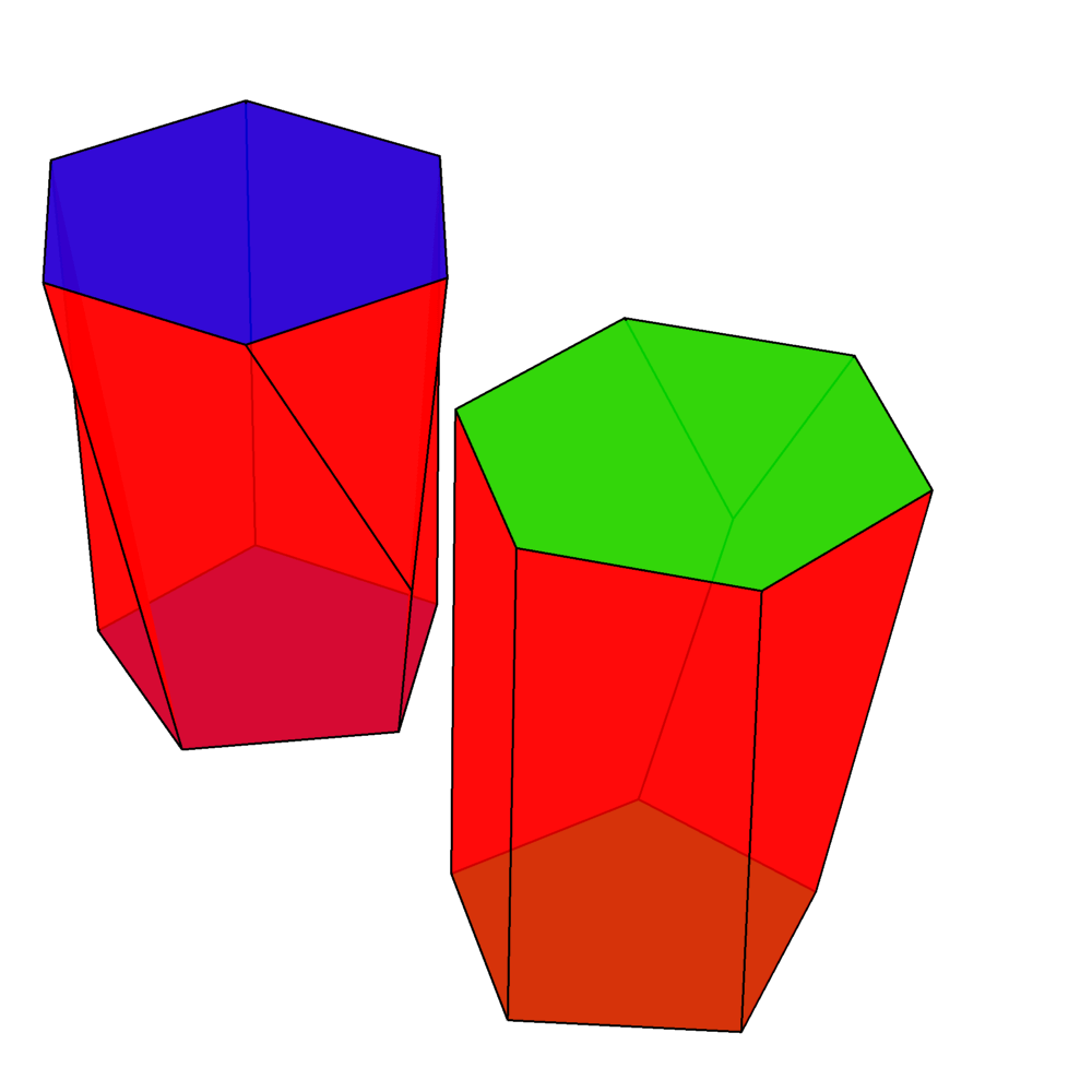
The family of shapes satisfying these conditions requires:
- Different numbers of vertices in the bases
- An additional vertex between the bases
- A Y-junction
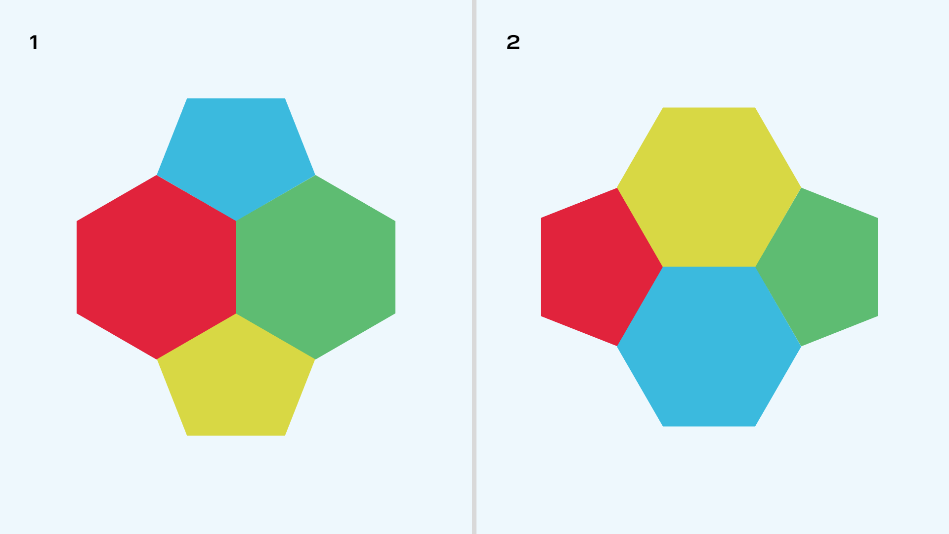
Interesting effect: Four scutoids joined so that two have hexagons on the bottom and two have hexagons on top create a situation where the neighbors at the bottom don't match the neighbors at the top.
Why the Scutoid Has This Particular Shape
The problem: Flat shapes do poorly at covering curved surfaces (think of a soccer ball or a globe unwrapped into a flat map).
With epithelium it's even harder: It sits at the boundary between the inside and outside of organs. Organs can have arbitrary shapes, the epithelium is three-dimensional, and the difference between outer and inner layers creates complexity.
The solution: Biologists used Voronoi diagrams for modeling.
Voronoi Diagrams
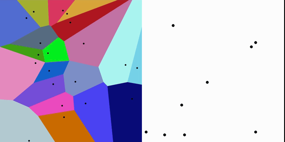
A Voronoi diagram is a geometric tool that divides space into regions closest to a given set of points.
Applications:
- Mathematics, biology, geography, physics, computer graphics, robotics
- Map generation in video games
- Determining nearest services (pharmacies, hospitals, schools)
Principle: Special points (seeds) create regions where every point inside is closer to its own seed than to any other.
Applying This to Cells
The two-sided structure of a cell:
- Apical side — faces the inside of the organ
- Basal side — faces outward
Modeling:
- Seeds are set as "centers" of cells
- A cell's shape is the region of points closest to its seed
- Modeling is done separately for the apical and basal sides (the results differ)
How the model develops:
- On flat tissue, cells take the shape of prisms (top = bottom)
- On curved tissue, top and bottom differ — requiring connection of different polygons
Why nature chose the scutoid: It is the energetically favorable shape that requires fewer deformations, better distributes mechanical stress, and leaves no gaps.
Distribution of Cell Types
The number of scutoids is proportional to the ratio of the radii of the surface areas of the layers:
- Ratio = 1: apical and basal surfaces are the same — cells are prism-shaped
- Ratio approximately 1: small differences — truncated pyramids
- Ratio much greater than 1: large differences — exclusively scutoids
- Intermediate cases: mixed packing
Future prospects: This model enables growing proper artificial organs and identifying pathologies.
And Finally
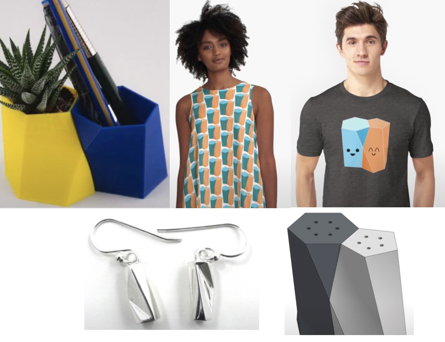
The discovery of the scutoid went viral on the internet. People created:
- 3D models for printing (available on Thingiverse)
- Themed clothing and household items
- A salt and pepper shaker model made from scutoids (link)
The original research paper was published in Nature Communications.