Rescuing the JEOL JEM-6A Electron Microscope
The story of saving a 1965 JEOL JEM-6A transmission electron microscope from a scrapyard fate — from discovering it on a classifieds site to transporting the 1.5-ton instrument to the Moscow Polytechnic Museum for preservation.
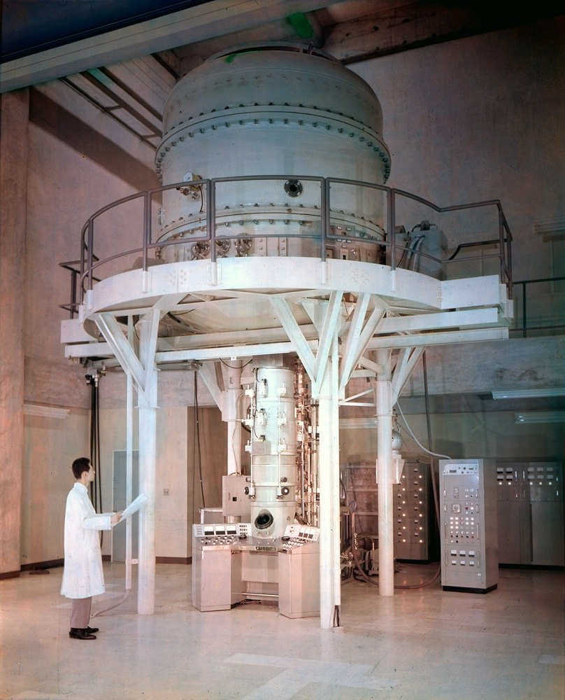
This is the story of how a 60-year-old Japanese electron microscope was saved from the scrap heap and delivered to one of Russia's most prestigious museums. It involves classified Soviet institutes, 160 liters of mineral oil, a 1.5-ton instrument, and a 400-kilometer journey through spring weather.
A Brief History of JEOL
JEOL — Japan Electronics Optics Laboratory — was founded in 1949 in Tokyo. The company began manufacturing transmission electron microscopes and quickly became one of the world's leading producers of scientific instruments. By 1955, JEOL was exporting electron microscopes globally, including to the Soviet Union.
The specific microscope in our story — a JEM-6A model — was acquired by the Nizhny Novgorod Institute of Technology (NIITOP) in September 1965. The institute used it for developing microelectronics manufacturing equipment, and work logs documenting decades of experimental procedures survived alongside the instrument.
How Electron Microscopes Work
To understand what makes this instrument special, a brief primer on electron microscopy is needed. Optical microscopes are limited by the wavelength of visible light — they can resolve details down to roughly 300-400 nanometers, but no further. Electron microscopes bypass this limitation by using electron beams instead of light, achieving resolutions down to 0.1 nanometers.
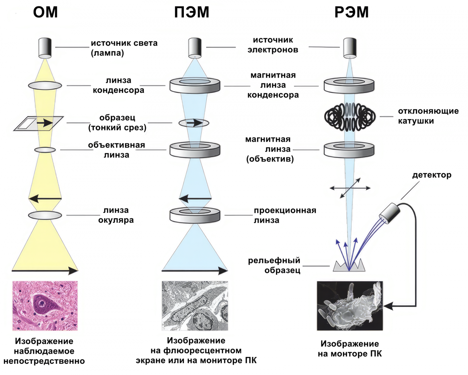
Two primary types exist: transmission electron microscopes (TEM), which pass electrons through ultra-thin samples, and scanning electron microscopes (SEM), which scan a focused beam across the sample surface. The JEM-6A is a TEM, operating at acceleration voltages of 50, 80, or 100 kilovolts.
During the 1960s, the race for ever-higher voltages led to impressive but impractical instruments. JEOL built the JEM-1000 in 1966, a megavolt electron microscope requiring an entire building to house. These supravoltage instruments ultimately proved economically impractical — beyond 300-500 kV, the X-ray generation and power consumption made operation prohibitively expensive.
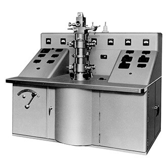
The JEM-6A: Technical Specifications
The JEM-6A was a serious research instrument with impressive capabilities for its era:
- Guaranteed resolution: 1.2 nm (optimal: 0.8 nm)
- Magnification range: 600x to 200,000x
- Acceleration voltage: Selectable 50, 80, or 100 kV
- Power consumption: 3-phase, 240V, 50Hz, 4.5 kVA
- Dimensions: 2,255 x 1,810 x 743 mm
- Mass: 1,480 kg
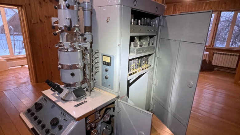
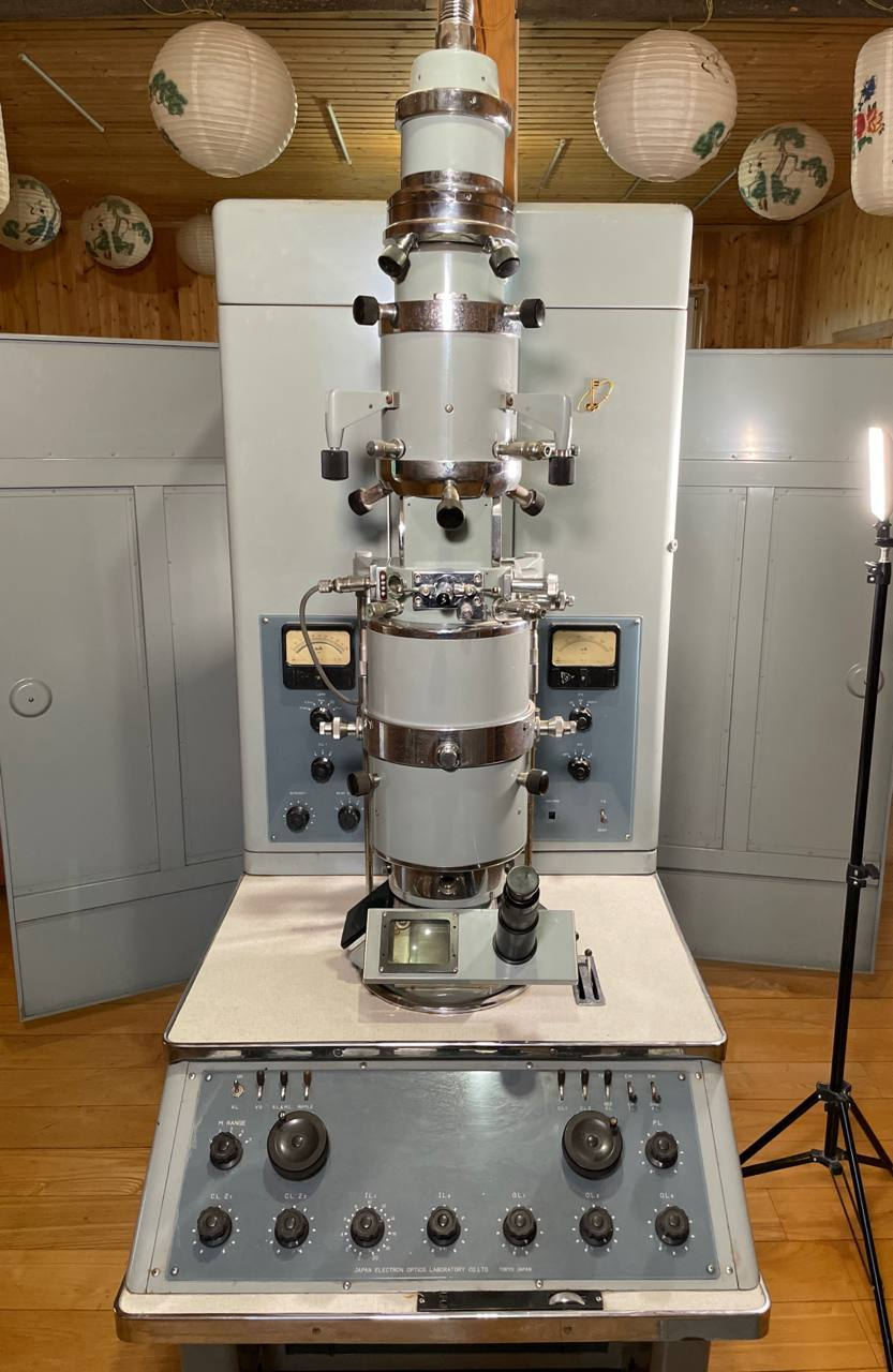
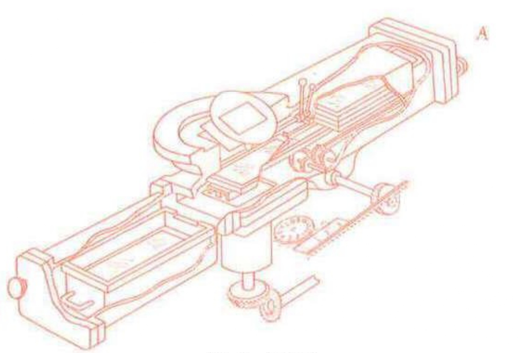
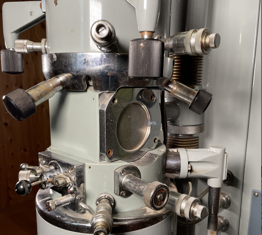
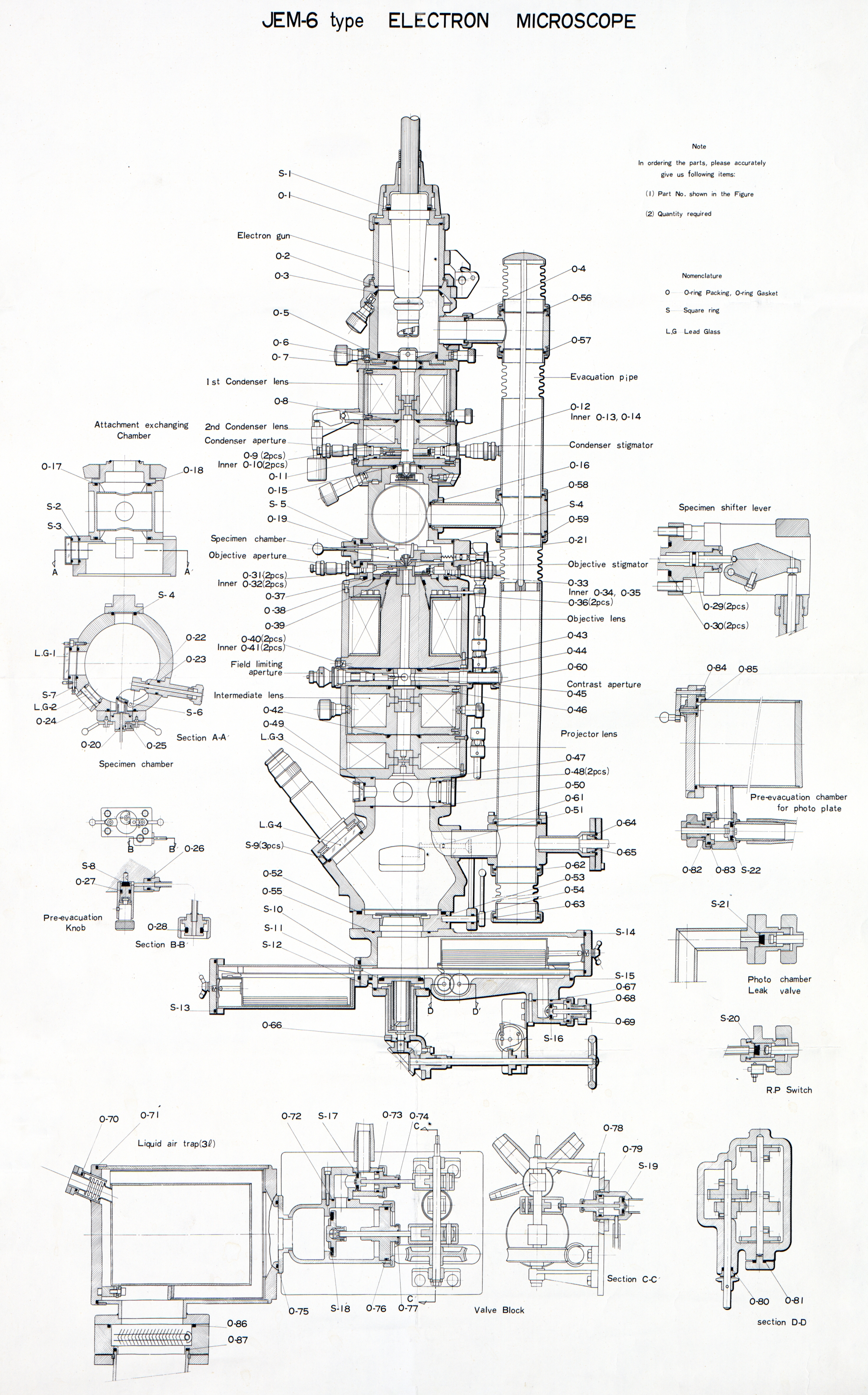
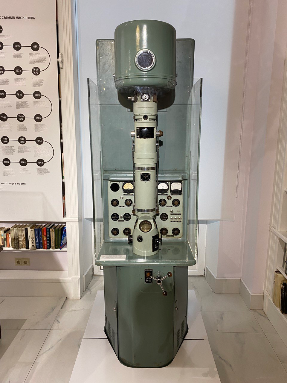
Inside the Electron Column
The electron column is the heart of any TEM, and the JEM-6A's column is a masterpiece of precision engineering.
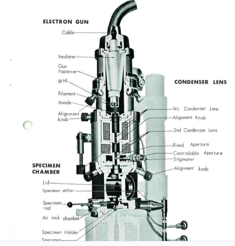
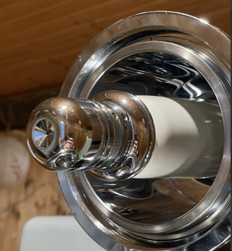
Electron Gun: At the top of the column sits the electron gun, generating a beam of 100 keV electrons from a tungsten cathode. The cathode is heated until electrons are emitted thermionically, then accelerated through a high-voltage potential.
Condenser Lenses: Two electromagnetic condenser lenses focus and control the beam before it reaches the specimen. These lenses use precisely wound coils to create magnetic fields that bend electron trajectories, functioning analogously to glass lenses in optical systems.
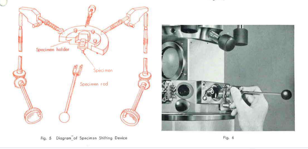
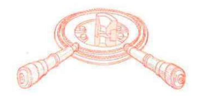
Specimen Stage: The specimen stage supports samples that must be thinner than 100 nanometers — thin enough for electrons to pass through. The stage provides mechanical positioning and can heat samples to +1,000 degrees Celsius or cool them to -140 degrees Celsius for studying materials under extreme conditions.
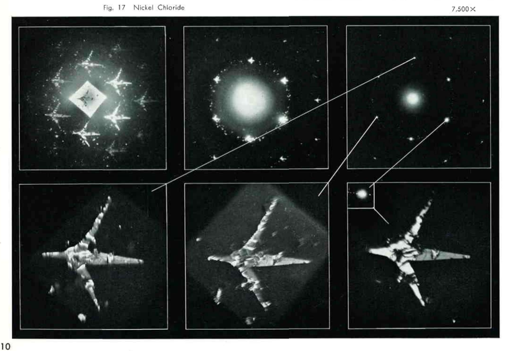
Projection Lenses: Below the specimen, three projection lenses — objective, intermediate, and projective — magnify the transmitted electron image and project it onto the viewing screen. Different lens configurations enable multiple imaging modes, including bright-field, dark-field, and electron diffraction.
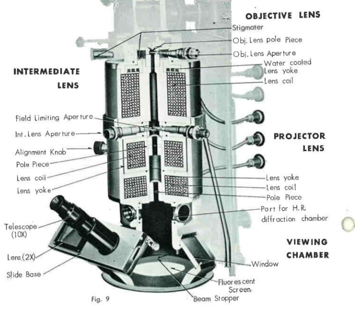
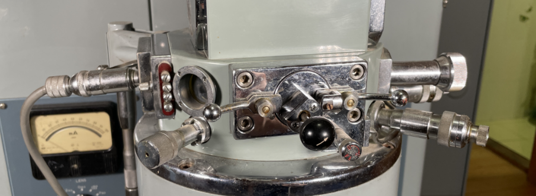
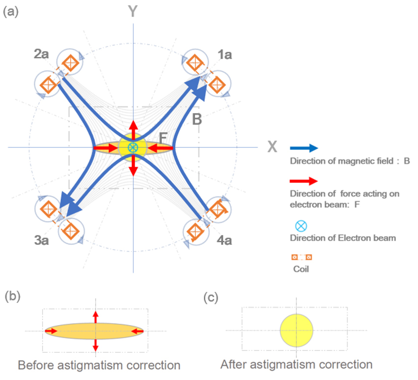
Stigmators: The JEM-6A uses mechanical stigmators — an unusual feature compared to modern electromagnetic designs. These employ ferromagnetic plates that are physically adjusted to compensate for beam asymmetries, ensuring the electron beam maintains a perfectly round cross-section.
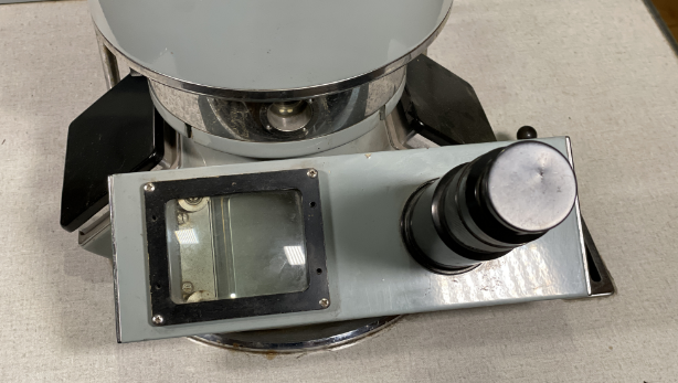
Viewing System: At the column base, a fluorescent screen converts the electron image into visible light. Operators view the image through binocular telescopes mounted at the screen level. For permanent records, an internal vacuum-sealed film magazine holds up to 24 photographic plates, mechanically indexed via lever mechanisms.
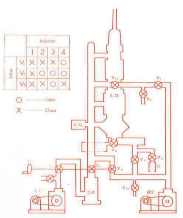
The Vacuum System
Electron microscopes require high vacuum — electrons scatter off air molecules, so the column must be evacuated to pressures of 10 to the minus 5th or 10 to the minus 6th Torr. The JEM-6A achieves this through a two-stage pumping system: a rotary vane fore-pump creates initial vacuum, then a diffusion pump (using boiling oil to trap gas molecules) achieves the final high vacuum.
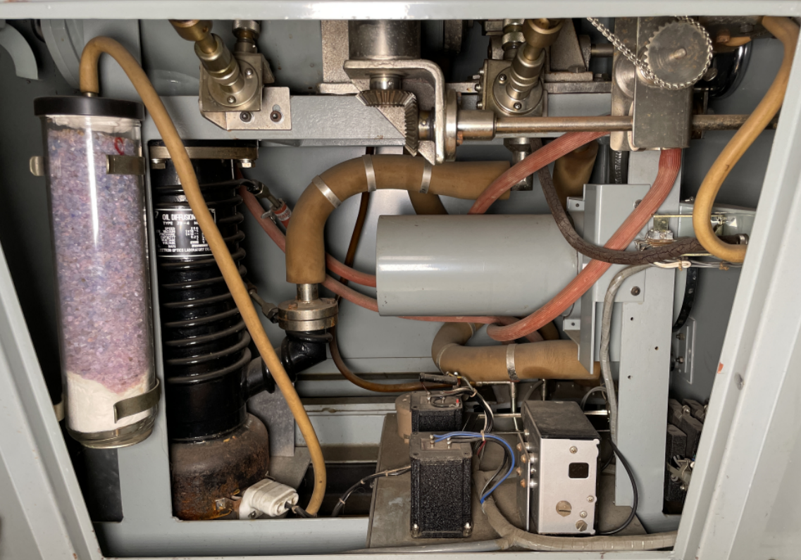
Two bellows vibration isolators flank the exhaust line, preventing pump vibrations from reaching the delicate electron column. This is critical — at 200,000x magnification, even microscopic vibrations would blur the image.

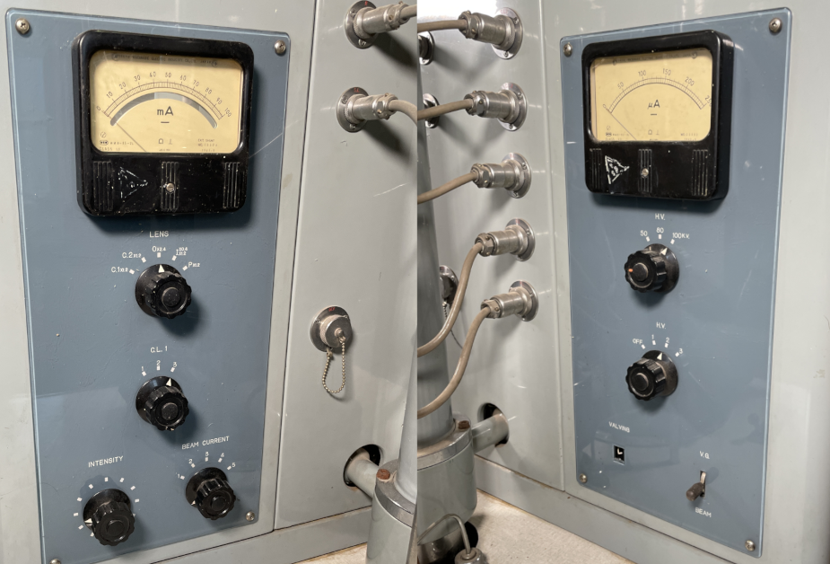
Control and Power Systems
Operating the JEM-6A is a manual affair. The control panel features rows of toggle switches that must be activated in a specific sequence. Initial startup requires a 20-minute warm-up period for the diffusion pump; full column evacuation takes an additional 5 minutes.
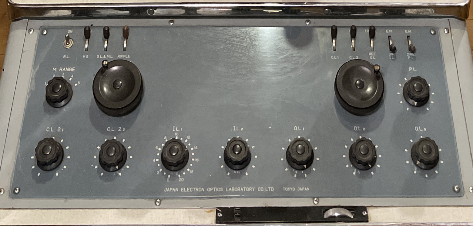
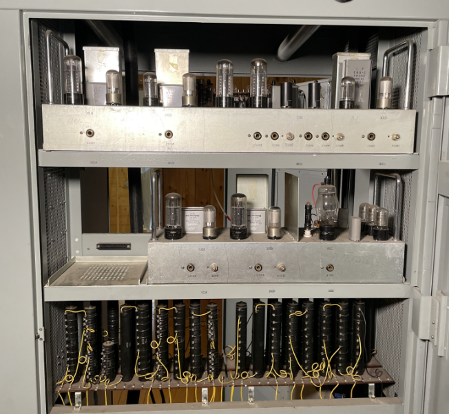
Two analog needle gauges provide essential monitoring: the left gauge displays magnetic coil current (controlling lens strength and thus magnification), while the right shows cathode voltage. All magnetic lens coils are powered by lamp-based (vacuum tube) power supplies — a purely analog system with no digital components whatsoever.
The Soviet Context
It is worth noting that the Soviet Union produced its own electron microscopes. The EM-3 was the first Soviet serial production TEM. However, Japanese instruments like the JEM-6A were highly prized for their superior optics and reliability. The fact that NIITOP acquired a JEOL instrument during the Cold War speaks to both the quality of Japanese manufacturing and the pragmatism of Soviet research institutions.
The Rescue Operation
Discovery (December 2024): I discovered the microscope listed on Avito (Russia's largest classifieds platform) — remarkably, within walking distance of my workplace in Nizhny Novgorod. Despite my personal interest in acquiring it, I recognized this was a museum-grade artifact at imminent risk of being scrapped. I contacted Alexey Butyrin (@BootSector) about the possibility of acquisition by the Moscow Polytechnic Museum.
Museum Approval: The museum curators agreed. Olga Tikhomirova, the microscopy department curator, traveled to Nizhny Novgorod for inspection despite terrible spring weather — snow and ice on the roads in April. She was particularly excited about the work logs that documented decades of experimental procedures. The microscope had been instrumental in developing microelectronics manufacturing equipment at NIITOP.
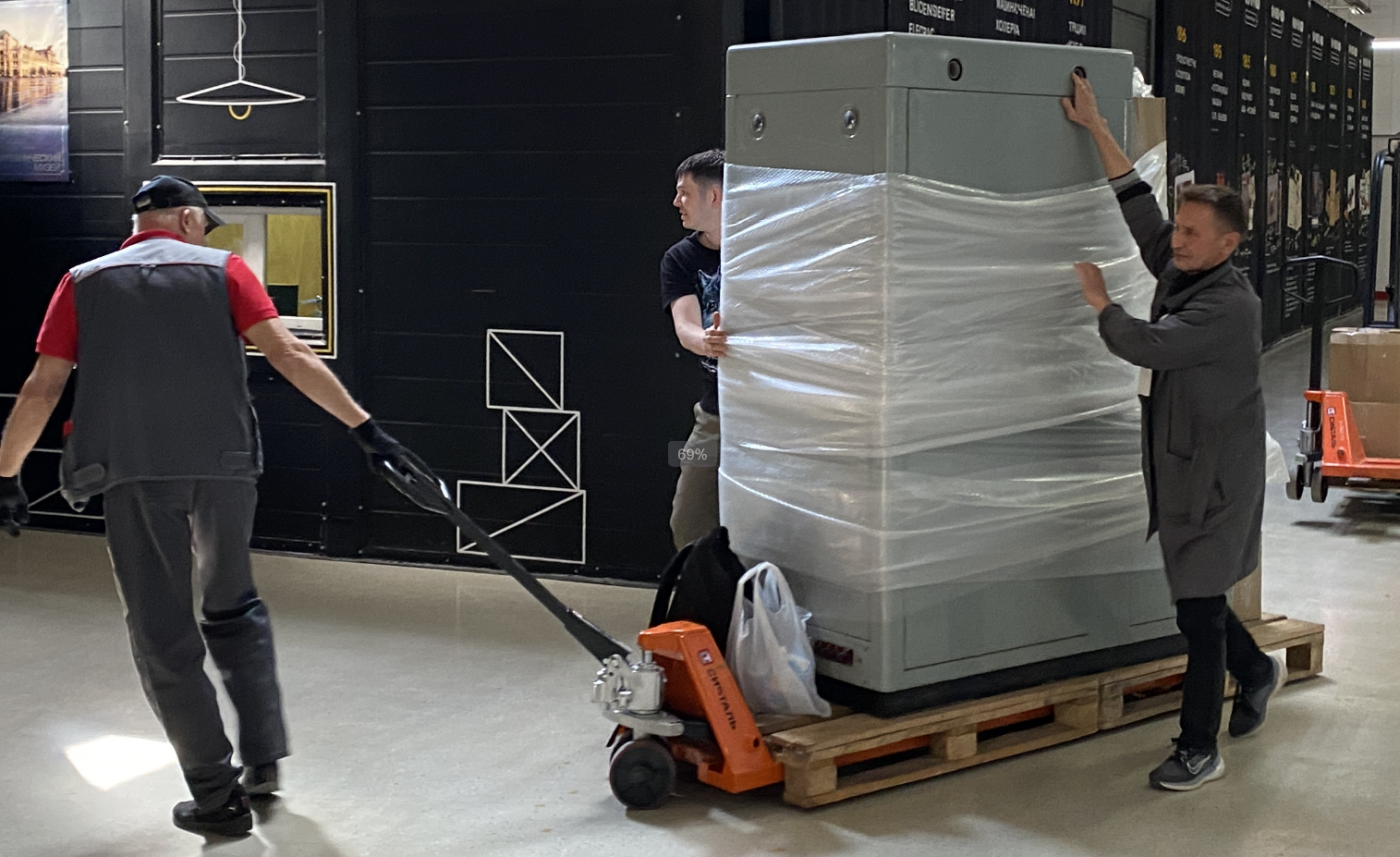
Preparation (May 2025): Moving a 1.5-ton electron microscope is not trivial. First, the transformer had to be drained of 160 liters of mineral oil — a modern petroleum product with a distinctive kerosene scent. The electron column head was removed and vacuum tube lamps were carefully packed separately to prevent damage during transit.
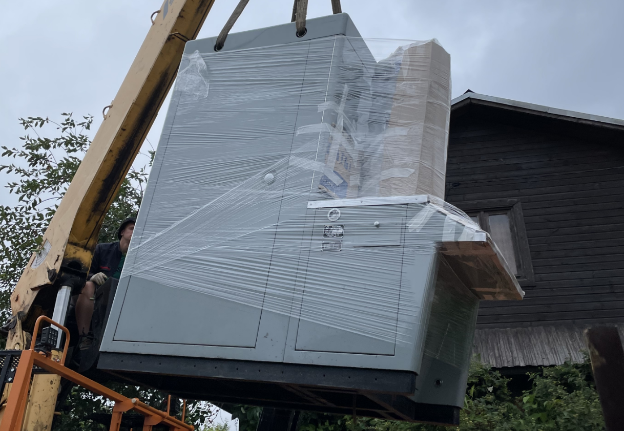
Transport: Professional movers loaded the instrument onto a specialized low-platform cart, then into a van. The 400-kilometer journey from Nizhny Novgorod to Moscow took approximately six hours.
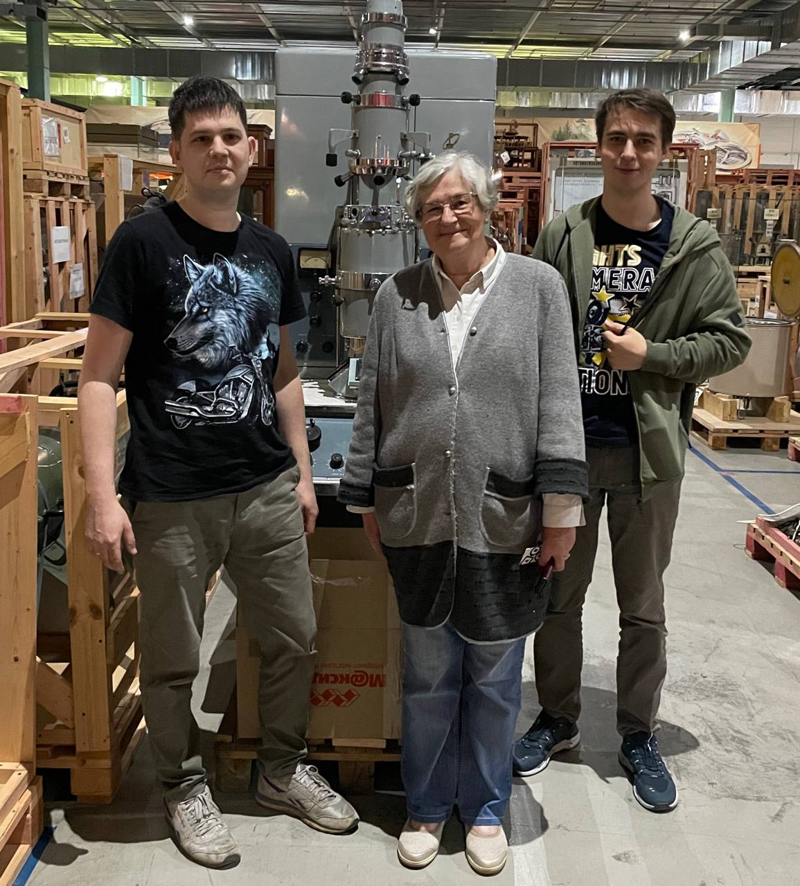
Installation: The microscope was installed in the Polytechnic Museum's open-access storage facility near Tekstilshchiki metro station in Moscow. This facility allows visitors to see museum artifacts that are not currently on main exhibition display.
Conclusion
Six months of anxiety — but the microscope is saved. The museum will not operate it (the complexities of maintaining a 60-year-old high-vacuum instrument with vacuum tube electronics make that impractical), but preservation for future generations is assured.
I am grateful to the museum staff — Alexey Butyrin and Olga Tikhomirova — for their professional support and boundless enthusiasm. Beyond the museum-allocated transport funds, personal contributions covered acquisition costs. It was a worthwhile investment for preserving an exceptional specimen of engineering artistry for succeeding generations.
The JEOL JEM-6A represents a pivotal moment in scientific instrumentation — when electron microscopy transitioned from exotic laboratory curiosity to essential research tool. This particular instrument witnessed decades of Soviet microelectronics development, and now it will continue to tell that story from behind museum glass.